![[Chronicle]](/images/sidebar_header_oct06.gif)
Medical, physical scientists team up to improve PET technology
By Steve KoppesNews Office
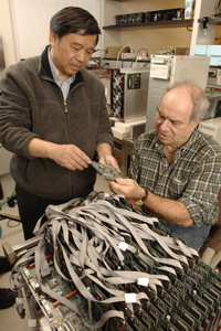
Photo by Lloyd DeGrane |
|
An international collaboration of medical and high-energy physicists will meet at a Thursday, March 27 workshop on the University campus to further develop a common technology that would serve dramatically different purposes.
The instrumentation could prove vital in both detecting disease and for making discoveries about the universe on the smallest of scales. “Modern electronics allow things that you couldn’t even think of 10 or 15 years ago,” said Henry Frisch, Professor in Physics and the College.
The collaboration consists of Frisch, Chin-Tu Chen, Associate Professor in Radiology, Chien-Min Kao, Assistant Professor in Radiology, and other scientists and engineers at the University, Argonne National Laboratory, Fermi National Accelerator Laboratory, Lawrence Berkeley National Laboratory, the Stanford Linear Accelerator, the University of Hawaii and the French Atomic Energy Commission.
If their work comes to fruition in the next few years, Positron Emission Tomography will be able to detect cancerous tumors at an earlier, more curable stage. And particle detectors in high-energy accelerators will be able to pin down the identities of more of their quarry.
At the workshop, the collaboration will determine the specifications for their system’s electronics. The meeting is partly funded by the French Embassy and the University’s France Chicago Center.
Frisch and Chen share a desire to more precisely measure the velocity of subatomic particles using an emerging technique called “time-of-flight positron emission tomography.” This technique can provide a positional measurement that is lacking in conventional PET technology—the kind widely used in medical imaging.
High-energy physicists, meanwhile, could use the technique to help identify many of the currently anonymous particles produced in their accelerator experiments.
“For the bulk of the particles that are made, we only know that a charged particle was created in the high-energy collision. We don’t know what kind it is,” Frisch said. The most common types of charged particles differ only in their quark content.
“It’s important because if you can identify the quark content of the particles, then you can look for very specific processes that are rare or forbidden in the ‘Standard Model’ that is the basis of our present understanding,” Frisch said.
Shared resources and expertise will be the key to their success, said Frisch and Chen. Physics students work in Chen’s PET laboratory, helping transfer the Enrico Fermi Institute’s capabilities in high-speed, large-scale electronic systems to the needs of medical imaging.
Frisch and Chen, along with Simon Swordy, Director of the Enrico Fermi Institute and the James Franck Professor in Physics and the College, also recently shared the cost of an ultra-fast Tektronix sampling digital oscilloscope, with further support from the Office of the Vice President for Research and for National Laboratories.
The oscilloscope has enabled them to compile a library of the types of signals generated by two different particle detectors. This allows them to determine how well certain kinds of electronics can measure the velocity and position of the particles.
“What people had done in the past was to assume some shape from the pulse and then simulate what the system would do,” Frisch said. But he and Chen’s team are now systematically running simulations based on real data. “We can ask how well a given system would perform without having to build it. And once you have it the way you want it, then you go and build the electronics that does that,” Frisch said.
In conventional PET scans, patients receive a dose of short-lived radioactive material that emits positrons. The PET scanner then detects the photons released when the positrons annihilate with electrons in the body. This approach generates millions of signals, including background noise that requires intense computational analysis to filter out. Furthermore, the signal locations can only be determined along the direction parallel to the detector face.
Time-of-flight data permits the determination of signal locations in a right-angle direction to the detector face as well. The capability of time-of-flight PET systems is assessed in terms of picoseconds. A photon of light can travel approximately one inch in 100 picoseconds.
Frisch’s goal is to achieve a resolution of one picosecond. Chen, who is measuring different types of events, would be satisfied with 30 picoseconds. This would provide improved PET scanner resolution in both directions and eliminate the need for expensive computational filtering of the background noise.
In their first simulations, the strategies that Chen and his associates tested had achieved a resolution of 100 picoseconds.
“That was pretty good. That was our first try,” Chen said. “We can do a lot better using an improved method for analyzing the data.” Kao presented the data last year at a meeting of the Institute of Electrical and Electronics Engineers in Hawaii.
“No one has this kind of collection so far,” Chen said of the pulse library. Testing the library against different particle detector configurations could cut down PET scan costs while also increasing image quality. “If we have very accurate digital measurements with modern computing and processing chips, we can actually cut down some of the manufacturing costs because we can get rid of some of the boards that were previously required,” he said.
Such are the potential payoffs when researchers pool their resources and expertise. The oscilloscope that Chen, Frisch and Swordy acquired is a shared instrument that will be available to other scientists in the Physical and Biological Sciences divisions.
“It will be used for high-energy physics, medical imaging and possibly astrophysics as well,” Chen said.